- Home
- Image Gallery
Select an application or technique:
Latest Images
High-Speed Imaging
AFM / OT and Advanced Optics
Life Sciences
Polymers
Nanoscience
Electrical and Magnetic
Cell Mechanics and Adhesion
Single Molecule Force Spectroscopy
Nanomanipulation and Lithography
Raman, TERS and SNOM
Atomic Force MicroscopyAutomated Force SpectroscopyOptical TweezersCellular Adhesion / Cytomechanics
AFM / OT and Advanced Optics
Superresolution Microscopy (STED, STORM, PALM)
-
 NanoWizard® BioScience AFM
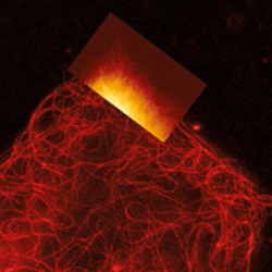
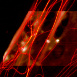
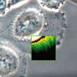
Living A549 cells - Correlative AFM and STED -
 NanoWizard® BioScience AFM
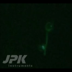
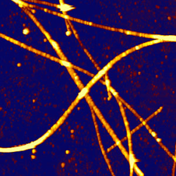
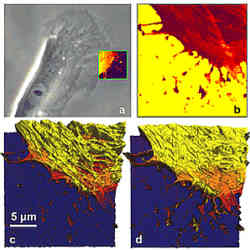
VIDEO - Real-time rapture of microtubules - Correlative AFM and STED measurements -
 NanoWizard® BioScience AFM
VIDEO - Real-time bending of microtubules - Correlative AFM and STED measurements -
 NanoWizard® BioScience AFM
VIDEO - Stimulation of living fibroblast cells - Correlative AFM and STED measurements -
 NanoWizard® BioScience AFM
Nanoruler - AFM and STED -
 NanoWizard® BioScience AFM
Living A549 cells - Simultaneous AFM and STED -
 NanoWizard® BioScience AFM
Simultaneous AFM and STED of Living fibroblasts - Actin Filament Imaging -
 NanoWizard® BioScience AFM
Simultaneous AFM and STED of Living fibroblasts -
 NanoWizard® BioScience AFM
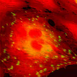
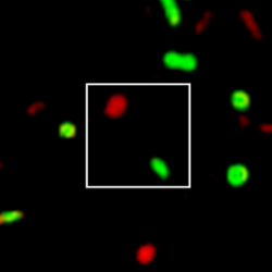
HeLa cell in buffer - AFM with STORM
Confocal Microscopy, FCS, FLIM, TIRF, IRM
-
 NanoWizard® NanoOptics A AFM
Simultaneous AFM and FLIM measurements -
 NanoWizard® BioScience AFM
Cell/particle interaction - AFM with confocal microscopy -
 NanoTracker™ 2
Recording of a confocal z-stack -
 NanoTracker™ 2
Confocal scanning combined with optical particle manipulation -
 NanoWizard® BioScience AFM
Lipid bilayer - AFM with confocal microscopy -
 NanoWizard® BioScience AFM
MDCK cells - AFM with confocal microscopy -
 NanoWizard® Sense AFM
SAOS cells - AFM with confocal microscopy
Fluorescence Microscopy
-
 NanoWizard® BioScience AFM
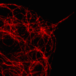
Rad51 proteins bound to DNA - AFM with fluorescence microscopy -
 NanoWizard® BioScience AFM
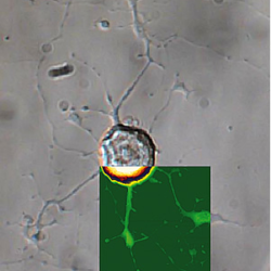
Living CHO cells - AFM with fluorescence microscopy -
 NanoTracker™ 2
Fluorescence filter change during trap manipulation -
 NanoTracker™ 2
Fluorescent microtubule manipulation -
 NanoWizard® NanoScience AFM
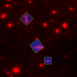
Hexaphenyl nanofibers - AFM with fluorescence microscopy -
 NanoWizard® NanoScience AFM
Fluorescent polymer spheres - AFM with fluorescence microscopy -
 NanoWizard® BioScience AFM
Ptk2 cells - AFM with fluorescence microscopy -
 NanoWizard® Sense AFM
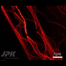
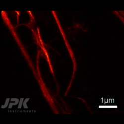
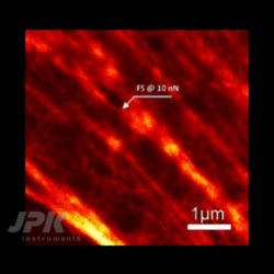
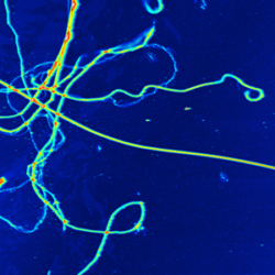
Microtubules – AFM with fluorescence -
 NanoWizard® BioScience AFM
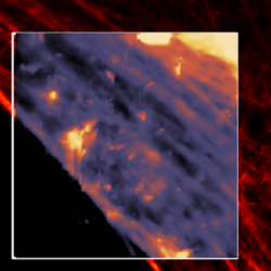
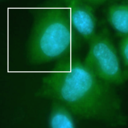
REF52 cells – AFM with fluorescence
Phase Contrast and DIC
-
 NanoWizard® BioScience AFM
Living Vero cells -
 NanoWizard® BioScience AFM
Living CHO cell -
 NanoWizard® BioScience AFM
CHO cell - AFM with phase contrast -
 NanoWizard® BioScience AFM
Living fibroblast cell - AFM with phase contrast -
 NanoWizard® BioScience AFM
Living dorsal root ganglion cells - AFM with DIC -
 NanoWizard® BioScience AFM
Collagen – AFM with phase contrast -
 NanoWizard® NanoScience AFM
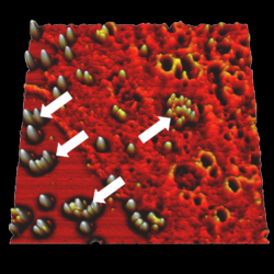
Polyelectrolyte shell - AFM with DIC -
 NanoWizard® Sense AFM
SAOS chondrocyte cells – AFM with phase contrast -
 NanoWizard® BioScience AFM
Living fibroblast cells – AFM with phase contrast and fluorescence
Upright Microscopy
-
 BioMAT™ Workstation
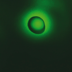
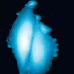
J-aggregates -
 BioMAT™ Workstation
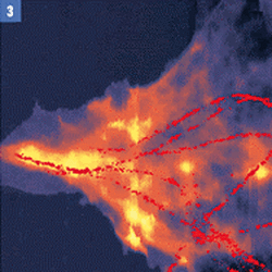
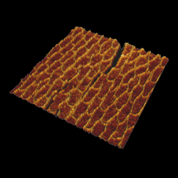
Paracoccus Seriniphilus bacteria -
 BioMAT™ Workstation
Living CHO on gold electrode -
 BioMAT™ Workstation
Cow tooth – etched - AFM with upright microscopy -
 BioMAT™ Workstation
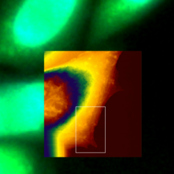
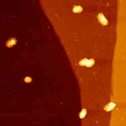
Bacteria on pyrite surface - AFM with upright fluorescence microscopy -
 BioMAT™ Workstation
Mouse cerebellum tissue - AFM with upright fluorescence microscopy