Correlative AFM and STED microscopy - gain deeper insights into your sample
Atomic Force Microscopy (AFM) is ideally suited for combining with super-resolution microscopy techniques such as Stimulation Depletion Emission (STED) microscopy. Both techniques address similar nanometer resolutions in biological samples and each comes with its own individual advantages. Whereas an AFM can manipulate and determine physical and chemical properties of surfaces, e.g. of a cell, a STED microscope can monitor biochemical processes, such as chemical recognition, inside the cell. The combination of these two techniques therefore delivers complementary data and provides a deeper insight into the specimen under investigation.
- Correlate topographical data with mechanical and bio-chemical cellular information
- Stimulate cells and tissues mechanically with AFM and monitor the cell response with STED in real-time
- Perform fast and high-resolution AFM/STED measurements with outstanding integration
The videos show (1) STED images of living human skin fibroblasts responding to mechanical stimuli being applied with a force of 10nN via AFM, and (2) real-time AFM manipulation of microtubules making them bend or (3) rupture (move the mouse over the movie and click the forward button to view all three videos).
 Perfect integration and powerful software enable easy data acquision
The integration of AFM with optical microscopy is our key area of expertise. JPK's NanoWizard® AFMs are compatible with the inverted microscopes of all major manufacturers and with advanced optical techniques such as STED and the microscopes from Abberior Instruments.
Our patented DirectOverlay™ software feature provides true correlative microscopy by enabling the perfect overlay of optical and AFM data with sub-diffraction limit precision.
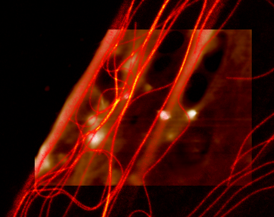
The image shows human skin fibroblasts microtubules, imaged with STED and overlaid with the AFM topography image (click on the image for details).
 JPK's intuitive QI™ mode makes AFM easy and delivers high resolution surface topography images while simultaneously determining mechanical characteristics of the surface such as elasticity and adhesion.
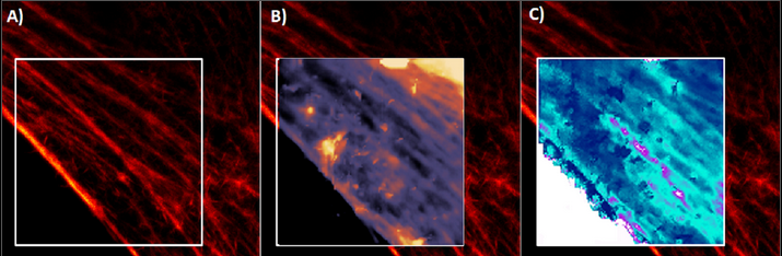
The images show (A) a STED image of the actin filaments of living fibroblast cells, (B) an AFM image of the topography and (C) the 3D topography image overlaid with Young’s Modulus (click on the images for details).
Further AFM/STED applications and technical details
- Check our Image Gallery for further examples of the combination of AFM with STED and other advanced optical techniques.
- Detailed information about instrumentation and applications can be found in our Application & Technical Notes section. In particular, the following notes deal with correlative AFM/STED:
 Quantitative and fast AFM co-localized with STED microscopy - towards living cells experiments Combining AFM with super-resolution STED microscopy
- The following webinar in the Webinars section gives an overview of both techniques and introduces applications in the fields of material research and life sciences (please register to watch the free recording):
JPK NanoWizard® AFMs in conjunction with Super-Resolution Abberior STED microscopes – towards live cell experiments
Selected AFM/STED user papers
- Desmoglein 2 regulates the intestinal epithelial barrier via p38 mitogen-activated protein kinase. Ungewiß et al., Scientific Reports volume 7, Article number: 6329 (2017)
- CLAFEM: Correlative light atomic force electron microscopy S. Janel et al., Methods in Cell Biology 140:165-185 (2017)
- Cellular level nanomanipulation using atomic force microscope aided with superresolution imaging.. Chacko et al., J Biomed Opt. 19(10):105003 (2014)
 The AFM-STED system incorporating JPK's NanoWizard® and Abberior's RESOLFT in the Lafont Group at BioImaging Center Lille (BICeL). Learn more
Correlative AFM/STED - Benefits
- Complementary information provides enhanced data
- Real-time monitoring of cell/tissue dynamics in response to mechanical stimuli
- Live tracking of changes in topography, adhesion, elasticity and other nanomechanical properties while simultaneously performing STED measurements
- Perfect overlay of STED and AFM data with sub-diffraction limit precision via DirectOverlay™
- True correlative multi-parameter microscopy by means of the intuitive QI™ mode
- Seemless integration of all NanoWizard® AFMs with STED microscopes such as the Expert Line and Compact Line from Abberior Instruments
Visit us at JPK headquarters in Berlin for demonstrations of simultaneous confocal, STED and AFM techniques using the combined Abberior STEDYCON and JPK NanoWizard® AFM setup.
![]()